Our research aims to understand molecular mediators of cellular mechanical behaviors, and their role in physiological functions and disease pathologies. Cellular mechanobiology—how cells deform, sense, and respond to mechanical cues—is critical in physiological and disease contexts.
We focus on three broad questions: (1) What are the molecular mediators of cellular mechanical behaviors, such as deformability, force generation, and mechanical adaptation? (2) How do soluble and mechanical cues regulate cellular mechanical behaviors? (3) How can we control cellular mechanical behaviors for translational applications, from enhancing chemotherapy response to accelerating tissue growth for food production? To address these questions, we use insights from physics and engineering to design novel experiments and analyze biological processes ranging from mechanotransduction to metastasis. Our unique and integrative approach uses the high throughput mechanotyping platform—invented and developed in the Rowat Lab—together with gene and protein expression data, bioinformatics analyses, and functional assays. With this new class of experiments, we aim to build a comprehensive map of the molecules and pathways that regulate cell mechanical behaviors in homeostasis and disease. While detailed knowledge of specific mechanical mediators has been established over the past decades, mapping the complete set of mechanical mediators and mechanosensing pathways—the ‘mechanome’—would enable us to predict and control cellular behaviors.
Here is a sampling of some of our current research projects:
Technology development for high throughput mechanobiology
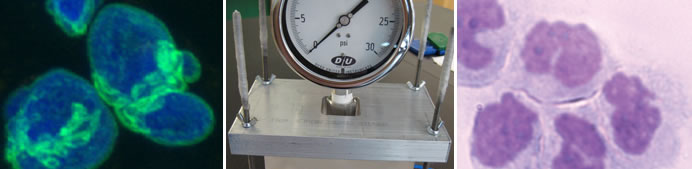
A major goal of our lab is to develop a systems-level mapping of the proteins and pathways that regulate cellular mechanical behaviors. To achieve this goal, small molecule screening would provide a powerful approach to identify previously unrecognized molecules that determine cellular mechanical phenotype or mechanotype. However, quantifying cell mechanical properties typically involves applying a physical force to cells one by one and measuring the resultant deformations, such as by micropipette aspiration or atomic force microscopy (AFM). Microfluidic methods enable faster measurements of single cell deformability but are still not compatible with high content assays that have revolutionized our understanding of biology, such as small molecule or shRNA libraries. To address this gap, our group invented High Throughput Filtration (HTF), which enables parallelized measurements of cell deformability in a format compatible with high throughput screening (Gill et al Lab Chip 2019); this builds on the prototype technology we previously reported (Qi et al npj Sci Reports 2015). We also invented a higher-throughput approach to obtain calibrated measurements of the elastic modulus of single cells, quantitative deformability cytometry (q-DC) (Nyberg et al Biophys J 2017). Our methods are contributing to research advances spearheaded by my own lab and ollaborators to discover novel mechanical mediators of cell behaviors in diverse contexts from cancer to tissue regeneration.
Regulation of cancer cell mechanical behaviors
The deformation of cancer cells through narrow spaces is essential in metastasis, and altered cellular deformability is associated with cell motility and invasion. To define how broadly conserved the relationship is between physical properties and motility across solid tumor cell types, my lab conducted a rigorous set of physical phenotyping studies across different types of cancer cell lines using both pharmacologic and genetic perturbations. Our findings reveal that cancer cells that are more invasive tend to be more deformable, but also identify contexts where more invasive cancer cells are stiffer than their less invasive counterparts (Kim et al J Cell Sci 2016; Nyberg et al Integrative Biol 2018; Nguyen et al Cell Mol Bioeng 2020). These findings are significant to advancing fundamental knowledge of cancer cell mechanobiology and also for applications of mechanotyping for cancer prognosis and drug discovery. My lab has also established novel mediators of cancer cell mechanical behaviors, including a β-adrenoceptor-actin-calcium axis that regulates cellular deformability, traction force generation, as well as invasion (Kim et al J Cell Sci 2016). We also contributed to the discovery of RhoA activation in the mechanical adaptation of circulating tumor cells, which promotes their survival (Moose et al Cell Reports 2020).
β-adrenergic (βAR) signaling regulation of epithelial and immune cell mechanical behaviors
Cells process information from diverse environmental stimuli, including both the stiffness of their environment as well as soluble compounds, such as stress hormones that are released into circulation in response to a physical or psychological threat. Yet little is known about how cells integrate these diverse forms of information from mechanical and soluble cues. Building on our discovery that βAR signaling increases the traction force generation by breast tumor cells, we are currently working to define how βAR activation impacts bidirectional cell-matrix interactions. To elucidate mechanisms of how βAR signaling increases traction forces in breast epithelial cells, we work with Dr. Parag Katira, San Diego State University, who has developed mechanistic models of cellular force generation. In collaboration with Drs. Sandra Orsulic and Steve Cole, UCLA, we are also working to test the hypothesis that β-blockers reduce cellular traction stresses and overall tumor stiffness to improve chemotherapy access using in vitro 3D tissue constructs as well as in vivo mouse models of breast cancer. These experiments are guided by analyses of gene expression signatures of patients in a prospective clinical trial of β-blockers in breast cancer by collaborator Dr. Erica Sloan, Monash.
Culturing muscle and adipose tissue for food production
Alternative production methods for animal protein are urgently needed to meet global demands and increase resiliency of our food system. The rapidly developing field of cultured meat—which addresses the challenge of growing muscle ex vivo by growing precursor cells harvested from animals in a bioreactor—has exciting potential to provide a sustainable alternative method for meat production with reduced environmental impact. But major advances in culture efficiency are needed to make this feasible and cost-effective. I hypothesize that the mechanical crosstalk between cells and scaffolds will be critical to achieve efficient production of muscle and fat tissues, which are key components of cultured meat. To test this hypothesis, we recently invented edible microcarrier scaffolds with tunable stiffness and surface topology that support the growth of muscle and adipose microtissue in a suspension culture. Importantly we can make these microcarrier scaffolds using food-grade ingredients with a scalable process. We are currently working to tune the mechanical properties of edible microcarrier scaffolds to increase the efficiency of culturing muscle and adipose tissue with reductions in costly media additives. Using high throughput screening approaches, we are also working to identify novel edible soluble cues that can accelerate lipid accumulation in cultured adipose tissue. Through collaboration with food scientists, we are working to define the sensory and nutrient properties of cultured meat produced with edible microcarriers; ultimately to achieve texture, mouthfeel, and protein content that is comparable—or even superior—to conventional meat, but at a lower cost and reduced environmental impact.
COLLABORATORS
Our collaborators include Beth Karlan (UCLA), Sandra Orsulic (UCLA), Erica Sloan (Monash/UCLA), Robert Damoiseaux (Molecular Shared Screening Resource/UCLA), Steve Cole (UCLA), Loren Fong (UCLA), Jenifer Fenton (Michigan State U),Jason Rowntree (Michigan State U), Andrea Garmyn (Michigan State U), Gale Strasburg (Michigan State U), Song Li (UCLA), Stephen Young (UCLA), and Parag Katira (San Diego State University).
|